Wednesday, November 10, 2010

Natural Variation in Thermotolerance Due to a cGMP pathway?


Most variations between conspecifics are often caused by simple differences in their enzymatic activities. These differences then in turn allow for evolution of the species. I came across this paper on variations in thermotolerance in fruit flies and locusts when one of the past PhD students, Gary, recently asked to me collect some additional data for one of his upcoming papers. To gain a better understanding in Gary’s field of study before the data collection, I read one of his prior publications from 2007.


In this paper, Gary and his colleagues manipulated the foraging gene (for), and its downstream pathways in locusts and fruit flies to examine their effects on the animals' ability to maintain neural and behavioural thermotolerance.
It has been illustrated that natural allelic variation in for gave rise to rover, forR, and sitter, forS in D. melanogaster larval foraging behaviours. Rovers are described as those larvae that moved more than sitters when feeding, and have higher PKG transcript levels and activities. The function of for in behaviour is said to be conserved across most organisms including ants, bees, nematodes, and mammals. In addition, for is also known to encode a cGMP-dependent protein kinase (PKG).


Thus, researchers hypothesized that the polymorphism in for can provide populations with natural variation in heat stress tolerance, and the variations in for and the differences in PKG levels could contribute to natural variations which might impact the evolution of thermotolerance in natural populations. .

This graph illustrated that forS2 had the highest thermotolerance
amongst the 3 variants, whereas the variant that has the highest
PKG activity, forR displayed the lowest heat-tolerance.
In this paper, researchers first examined whether different thermotolerance levels existed between Drosophila for variants. During these experiments, researchers increased temperature systematically and recorded the heat-tolerance failure point indicated by the cease in larval mouth hook movements. Three different types of for variants were used during this set of experiments. forR, forS, and forS2, which is a line of sitter mutant generated on a rover genetic background.

From these experiments, researchers found that forS2 illustrated a significantly higher failure temperature than forS and forR, and that forS and forS2 demonstrated a significantly higher synaptic transmission failure temperature than forR. This suggests that a lower PKG activity from the two forS variants could correspond to an increase in thermoprotection.

To confirm this hypothesis, researchers pharmacologically manipulated the PKG pathway by adding a cell-permeable PKG-specific inhibitor KT5823 and activator 8-Bromo-cGMP to Drosophila larvae. They found that the inhibition of the PKG pathway by KT5823 significantly increased the synaptic failure temperature in all three for variants, while PKG activation significantly decreased thermotolerance.

Researchers also explored the downstream effectors of PKG in thermotolerance by studying PP2A, which also affects K+ channels. PP2A is phosphorylated by PKG, which then in turn de-phosphorylates specific K+ channels. In this study, researchers noticed that a PP2A-specific inhibitor, Cantharidin increased the thermotolerance of synaptic transmission as strongly as the PKG inhibitor KT5823. Thus, they confirmed that there is a negative relationship between PKG activity and the thermotolerance in Drosophila larvae.
This graph illustrated that, heatshock, KT5823, and Cantharidin were able to increase the animals' thermotolerance while 8-Bromo cGMP decreased their heat tolerance by increasing their PKG activity. This graph suggested a negative relationship between PKG level and thermotolerance.
This graph illustrated that the PP2A inhibitor Cantharidin was able to
increase the animals' heat tolerance and decrease their time of
recovery. In addition, the effects of heatshock can be abolished by
an PKG activator 8-Bromo cGMP, of which the effects can be
abolished by the PP2A inhibitor, Cantharidin. This indicates a
sequential pathway starting with heatshock, PKG, and ending with
PP2A.
Moreover, researchers also studied the interaction between heatshock pretreatments and PKG pathways. In this set of experiments, by examining the effect of a PKG activator, 8-Bromo cGMP on heatshocked locusts, researchers concluded that there was no significant difference between the failure temperature and the recovery time in control locusts that were treated with and without 8-Bromo cGMP. However, the heatshock induced heat-tolerance was abolished by the application of 8-Bromo cGMP.

Furthermore, when compared with control locusts, the simultaneous addition of both PKG activator and the PP2A inhibitor in HS animals resulted in maximal thermotolerance of failure temperature and minimal recovery time. These outcomes indicated that PP2A is likely acting downstream from PKG and interacting with the same pathway affected by heatshock pretreatment.

In conclusion, this study determined that reduced PKG activity in forS resulted in greater thermotolerance, which can be replicated pharmacologically by using a PKG inhibitor, KT5823. In addition, the PP2A pathway is found to be downstream from the effects of heatshock and the PKG pathway, but upstream from the effects of K+ channels. These findings confirm the hypothesis that the polymorphism in for can indeed provide populations with natural variation in heat stress tolerance.

I really enjoyed this paper because of those very well designed experiments Gary et al. conducted in this study. However, one drawback of this publication was the redundancy of information in the figures, which made the result section seemed sloppy and confusing.
 
Reference: Dawson-Scully, K., Armstrong, G.A.B., Kent, C., Robertson, R.M., Sokolowski, M.B., 2007. Natural Variation in the Thermotolerance of Neural Function and Behaviror due to a cGMP-Dependent Protein Kinase. PLoS ONE 8:e773

Sunday, October 24, 2010

What about trafficking?

One other word that has frequently been mentioned in my blogs is 'trafficking'. Many people, even biologists might not understand what trafficking is or how it works. Thus, I think I will write another blog giving people some background of my research, in this case, trafficking.

Often, people would consider a negative connotation associated to the word 'trafficking'. For example, human trafficking, or drug trafficking. However in the scientific world, trafficking is not nearly as bad as its normal meaning. In fact, protein trafficking might have been an important feature that allows the development, and the evolution from prokaryotic to eukaryotic cells. There have been speculations that the protein trafficking systems would have to be evolved prior to the eukaryotic cells. Because simple cellular diffusion is often not sufficient as a method of substance transportation within eukaryotic cells, thus substance movements had to be archived via a specialized transport mechanism in eukaryotic cells rather than just diffusion.

Protein trafficking, or translocation involves rapid intracellular shuttlings of molecules packed in transportive vesicles. Cellular trafficking is often accomplished by following cellular tracks composed of cytoskeletonal proteins using motor proteins. In detail, some protein appeared to use different types of cytoskeleton tracks depending on the phase of the cell cycle. Translocation allows cells to store away unused proteins in vesicles, while rapidly mobilize and deploy them when a demand emerges. Though translocation, a cell greatly reduces its protein turn-over rates, which in turn, reduces the energy expenditure of a cell while performs all of its function at a timely fashion. It has been reported that the effects of translocation can be often seen within half an hour or less.

One of the important organelle used during trafficking is the Golgi apparatus. It plays two central roles in many cellular trafficking events; it is involved in protein synthesis and delivery, as well as internalizing molecular processes via endocytic. In more detail, brefeldin A, a chemical inhibits protein synthesis by blocking membrane trafficking from the endoplasmic reticulum to the Golgi apparatus proves that protein translocation is a necessary step in protein synthesis, as protein synthesis is often requires different organelles. In another study, it has also been found that besides protein synthesis, progression of murine oocyte maturation possibly also requires functional membrane trafficking.

Trafficking is a common phenomenon that has been observed across many species. For example, in humans, copper pumps are trafficked to the cell membrane from the E.R. in the presence of excessive cellular copper ions. Recently, trafficking has also been shown to be regulated by environmental cues. For example, in electric fishes, voltage-gated sodium channels are circadianly trafficked into the excitable membranes of electrogenic cells before conducting weak electric field for communication and navigation.

In conclusion, protein trafficking is not only a process that can greatly reduce the energy expenditure of a cell, while allowing all the cellular processes to run smoothly; it is also crucial in protein synthesis. Translocation might even be one of the features that allowed the evolution of higher organisms!

So, what exactly is a Na+/K+-ATPase

With all that many blogs thus far dedicated to this ATPase, I just realized maybe not everyone knows what a Na+/K+-ATPase is or what it does. Well, for those who are not as familiar with the Na+/K+-ATPase, here is a short background for ya.

The Na+/K+-ATPase, is a small, but nonetheless important membrane-bound active ion-transporter protein that is expressed in all multicellular organisms. This ATPase belongs the P-type ion-motive APTase family, in which also contains other H+, H+/K+, Ca2+, and heavy metal pumps. The Na+/K+-ATPase is a heterodimer containing alpha- and beta-subunits. The alpha-subunit is the catalytic subunit with a size of approximately 100kDa. In mammals, it has been noted the ATPase is composed of at least three a-subunits and three b-subunits. However, there is no evidence indicating the expression of different isoforms in insects, which is the one I'm studying. The active transport of ions across membranes by this ATPase is the greatest single energy-consuming process in most cells. Depending on the tissue type, the Na+/K+-ATPases consume between 5 – 40% of the cellular energy expenditure. Thus despite its small size, the Na+/K+-ATPase is a very important protein, especially in sensor/nervous/muscular cells where it is essential in conducting action potentials, and in renal cells where it regulates the reaborption of sodium.


 The key roles of this ATPase include, but not limited to regulating cell volume regulation, providing ion gradients that facilitate the movement of other solutes, and acting as a physiological regulator. Moreover, researchers have also demonstrated that in cardiac muscles, the Na+/K+-ATPase can even act as an indirect regulator of the myocardium contraction. In addition, the ion gradients provided by the ATPase across plasmalemmal membranes are also critical to cells, especially to those sensory, muscular and nervous cells.


Balanced Na+ and K+ ion-gradients across the neuronal membranes are essential for neurons to generate and conduct action potentials along their axons. The Na+/K+-ATPase assists neurons to sustain their Na+ and K+ homeostasis by shuffling Na+ and K+ ions across the plasma membranes. Through per ATP expenditure, this ion-transporter protein exports 3 Na+ ions out to the extracellular space for every 2 K+ ions imported into the cytoplasm. Due to the vital role of the Na+/K+-ATPase in sustaining action potentials, which is depended on the establishment of the Na+ and K+ homeostasis across the cellular membranes, the activity of this ATPase is targeted by multiple regulatory mechanisms to ensure its correct functioning. Failure in the regulatory mechanisms would often lead to deleterious effects.

 
To ensure its survival, a cell has overlapping mechanisms to failproof its regulations on the Na+/K+-ATPase. For example, the ATPase activity is enhanced by its substrates, or inhibited by cardiac glycosides. In addition, the Na+/K+-ATPase enzyme and substrate affinity also varies according to the cellular environment. Researchers have demonstrated that the addition of cytoskeletal proteins can increase the affinity of Na+/K+-ATPase for ATP. Furthermore, various hormones, such as insulin, and other signalling molecules can affect the activity of the protein positively or negatively. Moreover, the activity of the ATPase is also subject to regulation by reversible protein phosphorylation.

As you can sell, despite the relatively small size of the Na+/K+-ATPase, the rigorousity of the mechanisms that are regulating the Na+/K+-ATPase is undeniable. These mechanisms proved the importance of this protein and ensured that it is always being kept under its optimal condition to maintain the Na+ and K+ ion-gradients, a duty that is critical to an organism’s survival.

Thursday, October 21, 2010

The most expensive lunch...


Wednesday, October 20, 2010

Another Na+/K+-ATPase regulatory mechanism!

Recently, I have started writing the introduction of my thesis. Since my project is mostly involving Na+/K+-ATPase in locusts, I've planned to dedicate a substantial section of my introduction to talk about on the general background of the Na+/K+-ATPase. As I was looking through past studies for ATPase backgrounds, I've came across this paper that introduced another regulating mechanism on the Na+/K+-ATPase.

As people know, unlike tropical animals, sub-arctic organisms and poikilotherms such as insects, would have to overcome difficult winters annually. Through evolution, those animals have come up with different strategies to withstand the cold winter to ensure their survival. One of strategies the animals utilize is diapause (a hypometabolic state of arrested development). During this state, animals often suppress most of their metabolic processes and leaving on only the essential pathways to minimize ATP expenditure. As one of the most energy-expensive protein, the Na+/K+-ATPase which consumes 5 to 40% of a cell’s energy, has to be regulated during the diapause if the organism is to survive winter.
 
The researchers in this paper has found that the Na+/K+-ATPase activity can be reversibly suppressed by Protein Kinase A, C and G during winter to a point where only a fraction of the control activities were exhibited. Sequentially, the ATPase activity can be restored back to its normal state using a phosphate.

In this study, the researchers examined the Na+/K+-ATPase activity from the galls containing last instar larvae of Eurosta solidaginis. The researchers studied the ATPase activity during different winter months, as well as the effects of kinases on the ATPase. To study the ATPase winter activity, researchers assessed its activity in different sets of animals in October, November, January, February and April. They found that the ATPase activity was significantly lower in November and January with respect to October. Nonetheless, the ATPase activity recovered in February, and further enhaced in April.

This image illustrated that the Na+/K+-ATPase is siginificantly lowered in Nov., and Jan. with respect to Oct. However, the activity in restored in Feb., and further enhanced in April.
This figure illustrated that the Na+/K+-ATPase inhibition caused by PKA, PKC and PKG can be restored using a calf-intestinal alkaline phosphatease (CIAP).
In addition, when researchers studied the ATPase activity under the presence of PKA, PKC and PKG, they found that the kinases cause the ATPase activity to decrease to just 3 to 8% of the control values. However, the activity can be restored through the addition of a phosphatase.

This paper illustrated that during winter months, diapausal organisms can easily reduce their most ATP-consuming process, the Na+/K+-ATPase, by reversible phosphorylation via PKA, PKC and/or PKG. Once spring comes around, the animals can easily restore their ATPase activity via a phosphatase. The results from this study could suggest a biochemical pathway that could easily be utilized by other hibernating animals to conserve their ATP. In addition, the results from this paper also added to the general pool of knowledge of winter metabolic suppression.

Lastly, the parts I did not like about this experiments is that the researchers did not examine the activities of PKA, PKC and PKG during the winter months to prove that the activity decrease observed in winter months was actually due to the kinases. Furthermore, another area I was confused about this paper was that the effect that all the controls had a ‘non-1’ normalized value, which is normally impossible.

Reference: McMullen, D.C., Storey, K.B., 2008. Suppression of Na+/K+-ATPase activity by reversible phosphorylation over the winter in a freeze-tolerant insect. Journal of Insect Physiology 54:1023-1027

Sunday, October 3, 2010

Third time the charm?

This is my third trial on staining the Na+/K+-ATPase. The major problem I encountered in my first trial was from the sheath preventing the antibodies on penetrating and staining the ATPase. The major problem I encountered in my second trial was the poor tissue preparations from sectioning.

This time, after much research, I decided to use colleganse to dissolve the outer sheath enclosing the ganglia, and then stain the ATPase. However, the incubation length for colleganse could be tricky. Colleganse is a nonspecific enzyme that will digest will any collagen. This means initially the colleganse will digest the sheath surrounding the ganglia, but if you incubate your tissue in it for too long, the colleganse will then start digesting the cellular collagen and cause your tissue to lose its integrity. To determine the optimal colleganse incubation duration, I prepared three different samples each with a different incubation length, 6 hours, 12 hours, and 24 hours.

After much waiting, the results are finally in! It turns out that the 6-hour colleganse treatment was not sufficient enough to dissolve the outer sheath, but after the 24-hour treatment, the cell eventually loses its structure. As a result, the 12-hour treatment had the best compromise. Though, there is still dark area in the middle of the ganglion indicating and the antibody was still having trouble penetrating into the center...

This video shows the serial section images taken using the confocal microscope starting from the dorsal side of the ganglion.

With all the results in, I’m fairly happy with what I’m seeing, with the only thing I might want to change is trying to use an 18-hour colleganse treatment to see if that can improve my images. Now that I think I’ve got my staining protocol down, it’s time for some real results!

Friday, October 1, 2010

Will this paper finally be the one that will save my research project?

After my supervisor has seen the images from my second set of experiments, both of us went into a deep puzzle. He suggested that both of us should look deeper into literature that involved locust immunohistochemistry. One day, he sent me a paper that he thought I should ready and hopefully it help me to get the antibodies into the ganglia. Initially I was very skeptical at this paper because I didn't understand why would my supervisor send me a paper on locusts moulting rather than a paper on locusts' molecular biology or genetics which might actually involve immunostaining.

The paper he sent me involves studying the frontal ganglion (FP) and hormones involved during moulting.  The background for this paper is that unlike vertebrates, invertebrate growth requires molting. Successful moultings are essential for insect survival, which requires highly regulated hormonal and behavior coordination.
Past studies have indicated that at least four hormones, pre-ecdysis triggering hormone (PETH), ecdysis triggering hormone (ETH), eclosion hormone (EH) and crustacean cardioactive peptide (CCAP), are involved in the process of two phases of moulting, pre-ecdysis and ecdysis.

During pre-ecdysis, motor patterns are believed to loosen the old and new cuticles. However, the insect does not extricate itself from its old cuticle until ecdysis. The pre-ecdysis behaviours were triggered by the effects of PETH and ETH acting on the central nervous system (CNS). Centrally released EH is thought then triggers ecdysis behaviour, either directly or followed by a release of CCAP within the CNS in a cGMP-dependent manner. CCAP then triggers the motor activities necessary to complete ecdysial behaviour.

The researchers in this paper have previously described a novel central pattern generator (CPG) network situated in the locust FG, and the motor patterns it generates. In the desert locust, Schistocerca gregaria, FG neurones innervate foregut dilator muscles and play a critical role in the control of foregut motor patterns in
different physiological and behavioural states. The researchers have presented the FG as an important target for chemical modulation.

During moulting, the foregut and FG are involved in air-swallowing behaviour. By filling the gut with air, the larval locust can generate enough internal pressure on the body wall to eventually split open the old cuticle, and to then stretch and shape the new adult cuticle and wings after the old cuticle has been shed at ecdysis. Frontal ganglionectomy abolishes air-swallowing and results in difficulty or failure in eclosion and wing expansion. A role for the FG during moulting has been reported for a number of insect species.

In this study, the researchers used suction electrodes to examine the FG CPG as an unexplored target for ecdysis peptides. They also examined the density of CCAP using immunoblotting, and the location of CCAP and cGMP through immunohistochemistry. In their study, they identified ETH, and PETH as FG bursting frequency promoters, while EH as a FG bursting frequency inhibitor.
Figure A illustrated that ETH significantly increased the burst frequency in locusts in CPG. Figure B illustrated both ETH and PETH can increase locusts' burst frequencies, however, ETH was a significantly potent burst frequency promotor than PETH. Figure C illustrated that after the application of EH, locusts' burst frequency stops, but the inhibition is not permanent.
In addition, the researchers also identified CCAP as a potent modulator of the locust FG motor patterns. With applications of CCAP, frontal connectives (FC) and medial pharyngeal nerve (MPN) showed a significant increase in bursting frequency. The increased bursting frequency diminishes when CCAP is washed away with saline. In semi-intact preparations, CCAP also resulted in air bubbles in the crop, which is presumably the effect of air swallowing. Their results imply a novel role for this peptide in generating air swallowing behaviour during the early stages of ecdysis.

Figure A demonstrated that after CCAP application, there is a significant increase in burst frequency within the FC and MPN. However, the effects of CCAP can be diminished after washing the tissue. Figure B illustrated the air bubbles formed on top of the tissue after CCAP application as the result of air swallowing.


Last but not the less, from immunostaining researchers found that one to three CCAP-immunoreactive neurones in the tritocerebrum that extended through the FC nerve and gave rise to extensive arborization within the neuropil of the FG. Their immunostaning examined the variation in the number of CCAP immunoreactive axons in the FG neuropil at the different stages. In mid-larvae only one to two (mostly one) fibres were stained, whereas at the air-swallowing stage at least three afferent fibres were visible. As ecdysis progressed the number of stained axons then declined. A quantitative analysis concluded that the area of CCAP-immunoreactive neuropil increased during airswallowing and decreased at late ecdysis.

These series of immunostaining illustrates the number of CCAP-immunostaining reactive axons and the size of CCAP-immunostaining reactive neuropil in the the FG. These images demonstrated that the number of CCAP related axons increases in size as moulting occurs, but drops at late ecdysis, same logic applies to the size of CCAP related neuropil.
After all findings on regulators on locusts moulting, the part of this paper that might really help me in my project is found in their immunostaining section, when they stated '... (the tissues) were incubated in 0.5 – 1.0 mg/ml collagenase in PBSTX for 30 – 60 min at room temperature to aid in antibody penetration.' This is exactly what I wanted to read! Since my problem was that my antibodies wasn't entering the cells. This definitely gave me new energy on returning to my experiments again.


However, the part I didn't like in this paper was that many of the factors in the immunostaining had a relative large uncertainty in their incubation time. For example, as mention earlier, the tissues were incubated between 30 to 60 mins in collagenase, which might caused a difference by itself in staining results. Furthermore, this would mean I had to try the difference time frames during different incubation processes too to find an optimal time myself.

Ref:
Zilberstein, Y., Ewer, J., Ayali, A., 2006. Neuromodulation of the locust frontal ganglion during the moult: a novel role for insect ecdysis peptides. The Journal of Experimental Biology. 209:2911-2919.

Thursday, September 30, 2010

Trial 2

After I collected my thoughts back together and got over about my experiment did not work, I concluded that the reasons it failed could either be because 1) the antibodies would not penetrate through the sheath, or 2) the antibodies are not binding onto the ATPase (which I doubted and really hoped was not the case, because from the background research that was the only Na+/K+-ATPase antibody other researchers have used on insect Na+/K+-ATPase. Therefore, I didn't have much choice in terms of choosing antibodies). After discussing the problem and possible solutions with my supervisor, he suggested that I could use paraffin and sectioning to mount the tissues in wax and section the ganglia into slices to eliminate the problem created by the sheath. Therefore, if this experiment worked, then we can conclude that the first time my experiment didn't work was because the antibodies weren't able to penetrate the sheath and rule out the possibility that the antibodies are not binding on to the ATPase.

After I have my ganglia sectioned by a lab technician from another department, paraffined and stained, I'm ready for round 2 of confocal. This time the staining worked (the green fluorescence),  indicating that antibodies were able to enter the cell body and stained the ATPase. But as one problem is solved, another problem emerged. Because of the fragility and the size of a ganglion, which is roughly the size of a pen's tip, the ganglia were heavily damaged during the sectioning process and orientated in a random fashion. As a result, we were not able to identify the neurons within each ganglion and to make valid comparison between control and heat-shocked ganglia. Consequently, it appears that paraffin and sectioning also may not be the route to go about my experiment. Now, I will need to come up with some other method that does not involve sectioning but yet still allows the antibodies to penetrate through the sheath...

Sunday, September 26, 2010

Before the data collection...

If you are going to ask me to sum up my research project in one sentense, it would probably be something like this: using fluorescence antibody to study whether the location/trafficking of the Na+/K+-ATPases differ between a control locust and a locust that has received a heat-shock pre-treatment in their meta-thoracic ganglia (a pile of neurons enclosed by a layer of sheath). Sounds pretty easy you say? Yeah, that's what I thought too, before I started my experiments...

I've learned couple of things in the past year I've spend on doing my Master’s. For example, your experiment always take much longer than what your expected or as indicated on your protocol. But an even more important lesson I've learned was if this is the first time your lab doing a type of experiment, the chances are, it probably will not work at the first try;

My experiment sounded simple enough, dissect out the ganglia, incubate them with antibodies, and then study them through a confocal microscope; but the reality was far from that...

Trial 1:
Like a kid going to his first day of school, I was very excited about starting on my immunohistochemical experiments. I did my research and came up with a feasible protocol. I followed the protocol word to word, incubating the ganglion in antibodies  (red fluorescence) to stain the ATPases and DAPI  (blue fluorescence) to stain the cellular nuclei. Once the samples were ready, I just could not wait to see what they would look like under the confocal.
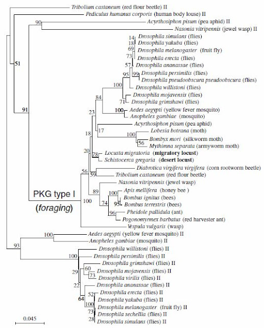
But to my least expected, this is what came up under the confocal... At first, I had no idea what was going on. But after my supervisor has looked at the images, we concluded only DAPI was small enough to penetrate the dense shealth that is enclosing the ganglia. Consequently, the sheath blocked out all the antibodies, resulting in only those visible cellular nuclei stained by DAPI. The red that we are seeing in our images were the visible antibodies that were the residues trapped inside of the ganglia trachia.

Well, I guess my first trial didn't work, looks like it's time for me to do more research and back to the drawing board again...

Saturday, September 25, 2010

The paper that layed the foundation of my staining protocal

During my background research, my supervisor sent me a paper for me to read, he said this paper will help me with my immunostaining protocol. The paper he sent me was conducted by researchers from Canada, Israel and Switzerland on locust foraging gene.

The background for this paper is that animals, including locusts display a astonishing genetic interaction with their environment to generate behavioral plasticity. In this study, the model animal researchers used were migratory locusts, Locusta migratoria, and desert locusts, Schistocerca gregaria. During their solitary-phase, those animals actively avoid contact with conspecifics and are relatively sedentary. However, they become more active, and start marching in bands of hoppers or flying in swarms in their gregarious phase, when their population density increases.


So far, numerous neurobiology studies have characterized the differences in locusts' sensory interneurons that faciliated this behavioral change. In contrast, little has been done to study the molecular and genetic contribution to this phenomenon.

Recently, there are a handful of other well-studied animals that have shed light on the genetic and  pathways that mediate environmental and behavior variations and plasticity. Recent studies have found that those foraging-related behaviors have been linked to the foraging gene (for) and a cGMP-dependent protein kinase (PKG). For example, it has been found in Drosophila melanogaster that allelic variation in for, for mRNA and PKG have been associated with a similar foraging phenomenon. Interesting, those with a rover allele (forR) exhibit a greater moving distance while feeding comparing to those with the sitter allele (forS).


In their study, researchers demonstrated that different allele variations were favoured under different environments; in a crowded population, the environment favours those with forR, while uncrowded population favours those with forS.


The researchers in this study cloned the locust for gene, placed it in an insect phylogeny of PKG, examined for expression in locust brain and measured the PKG activity. In this paper, the researchers confirmed that locusts grew up in different population-density colonies illustrated differences in development, coloration and pattern, neurophysiologies and behavior... ...

Researchers also found that the two species of locusts, L. migratoria, and S. gregaria, shared 90% pairwire identity and a 70% pairwise similarity in for gene when comparied with D. melanogaster. Further more, the for gene in locusts had the typical PKG domains, and two cGMP-binding domains. The researchers also compared locust's for sequences with all currently known insect PKG-encoding genes. They found that within the type I PKG cluster, a group formed by the dipterans and a group with all the social insects except the wasp Vespula vulgaris can be distinguished. The two locust sequences are the locust foraging genes and are part of the PKG type I cluster. 
This phylogenetic tree illustrated the phylogenetic analysis (Neighbour joining method) of the relationship of 44 PKG sequences spanning 30 insect species (variant sequences were discarded). Pairwise comparisons using 5,000 bootstrap replications were used to build the tree. The 2 locust species are in bold.
During the PKG activity assay between the two phases of locusts, researchers concluded that the PKG activity in gregarious locusts was significantly higher than that in solitary locusts. The differences in PKG activity are also sex specific, with higher PKG activity found in males. However, no interaction was found between sex and phases. This sex difference in PKG activity was found for both solitary gregarious locusts.


Through neruonal staining, the researchers also identified the locust for gene expression location within the locust brain.
This group of images illustrates the FOR expression patterns in the locust brain. A: Merged confocal image of frontal 3-mm optical sections of a locust brain showing the major brain neuropils (in green, mouse antibody). B: A distinct PKG-IR region is seen as a cluster of cell bodies in the anterior midline of the brain (double staining with anti-FOR in red and mouse antibody in green). C: Similar FOR expression patterns is seen in gregarious (Gre) or solitary-reared animals (Sol), in adult male (M), female (F), and larvae. Each scale bar represents 250 mm.
What really spiked my interested in this paper was the section of their method on immunocytochemistry and neuronal staining.


'Immunohistochemistry was as in Belay et al. (2007). Briefly, whole brains were dissected in PBS (0.1 M, pH 7.4), fixed in 4% paraformaldehyde, and blocked in 4% normal goat serum (Jackson ImmunoResearch, West Grove, PA) in 0.5% Triton X-100/PBS. The specific guinea pig antibody called anti-FOR, described in Belay et al. (2007), was used at 1:150; the neuropile marker mouse mAb nc82 was used at 1:20 (22, 23). Incubation was for 48 h at 41C. After incubation, brains were washed several times in 0.5% Triton X-100/PBS before adding a goat Cy2-conjugated anti-mouse and a Cy5-conjugated anti-guinea pig Ig (1:100, Jackson ImmunoResearch) for 24 h at 41C. For negative controls, brains were incubated in only secondary antibody, in the absence of primary antibody or in pre-absorbed anti-FOR serum. The specificity of the primary antibody, anti-FOR antibody generated in guinea pig, was measured by using Western blot immunodetection.'


What I really liked about this method section was that the steps for immunostaining was very clearly layed out, which I can copy and adjust for my experiment. The researchers also thoroughly tested the specificity of the primary antibody through Western blot immunodetection. In addition, they ensured the staining they were observing were not just background by omitting the primary antibodies in the negative control. However, the researchers did not indicate how long they fixed their tissues for, nor did they indicate the length for blocking in normal goat serum.

None the less, I got my immunohistochemistry sorted out in this paper, now it's time to fine tune the details to get everything work for my experiment!


Ref:
Lucas, C., Kornfein, R., Chakaborty, M., Schonfeld, J., Geva, N., Sokolowski, M.B., Ayali, A., 2010. The locust foraging gene. Archives of Insect Biochemistry and Physiology. 74:52-66.

Friday, September 24, 2010

The origin of my project

For every research project, there will always be a predecessor that led to the current project. Hopefully, there will also be a successor that follows the it. The paper that started my project was a paper published from our lab. In this paper, the researchers used the locust ventilatory central pattern generator (CPG) to study spread depression(SD)-like events. SD is closely associated with pathologies such as stroke, seizures, migraine and epilepsy. It is been characterized by a wave of electrohphysiological hyperactivity in the neurons followed by a wave inhibition.

In this experiment, neuronal hyperactivity is stimulated in locusts' ventilatory CPG by several stressors such as anoxia, ATP depletion, and hyperthermia. The neuronal acitivty is then monitored using  K+ sensitive microelectrodes to measured the extracellular K+ concentration in the metathoracic neuropile of the CPG. In addition, CPG output is monitored electromyographically from muscle 161 to investigate the role K+ in failure of neural circuit operation induced by those stressors.

In this study, through using K+ sensitive microelectrodes and electromyographic electrodes, researchers found the stress-induced ventilatory pattern failure is tightly correlated with a abrupt increase in the extracellular potassium concentration. This suggested that the failure of ventilation in response to different stressors (hyperthermia, anoxia, ATP depletion, Na+/K+ ATPase impairment, K+ injection) was associated with a disturbance of CNS ion homeostasis. This disturbance in CNS ion homeostasis also shares the characteristics of cortical SD and SD-like events in vertebrates.
These figured illustrated the tight correlation between the failure in ventilatory CPG and the abrupt increase in the extracellular potassium concentration. Noticeably, locusts were able to recovery their ventilatory motor pattern once they have restored their extracellular potassium homeostasis. Vent: ventilatory motor pattern monitored using and EMG electrode, [K+]o: extracellular potassium concentration monitored using the K+-sensitive microelectrode.

In addition, researchers found that heatshock delayed the onset of ventilatory pattern failure and shortened the time to restore Na+/K+ homeostasis and recovery of ventilatory pattern failure. The researchers found that this phenomenon is archived through an increased rate of extracellular potassium clearance by heatshock.

These two figure illustrated that HS decreased the rate of extracellular potassium accumulation and increased the rate of extracellular potassium clearance. This phenomenon hinted that after a HS pretreatment, locusts were better at restoring their potassium homeostasis.


However, when researchers studied the mechanism behind this increasesd clearance rate, they found that it was not due an increase in Na+/K+-ATPase enzymatic activity in heatshocked locusts. They measured the production of NADH spectrophotometrically at 340 nm. The Na+/K+-ATPase activity was assessed using a pyruvate kinase/lactate dehydrogenase assay. In this experiment, both Na+/K+-ATPase and pyruvate kinase/lactate dehydrogenase assay required ATP. The pyruvate kinase/lactate dehydrogenase assay produces NADH, and will compete for ATP with Na+/K+-ATPase. If the ATPase had a higher enzymatic activity, then it would utilize ATP at a faster rate, minimizing the amount of ATP is used for the pyruvate kinase/lactate dehydrogenase, in return, decreases NADH production

In addition, researches examined the cellular ATP level in heatshocked and control animals. The ATP level in heatshocked and control animals was quantified using a luciferin-luciferase reaction. In this reaction, ATP is consumed and light is emitted when firefly luciferase catalyzes the oxidation of D-luciferin. Since ATP is the limiting reagent, the the amount of ATP present is proportional to the light emitted, which is measured using an Lmax luminometer. Each measurement of light intensity was compared to a standard curve generated using known quantities of ATP prepared in water. The end result illustrated that there was not a significant difference in cellular ATP concentration between control and heatshocked animals. This indicated that the lower clearance rate observed in control locusts was not due to a lower level ATP, which could hinder their Na+/K+-ATPase function.

This figure illustrated that there was no significant difference in cellular ATP level between control and heatshocked locusts at the same time. However, a significance was observed during anoxic period for both control and heatshocked locusts.

This study concluded that when hyperthermic failure that was preconditioned by prior heat shock, it resulted in an induced-thermotolerance which was associated with an increase in the rate of clearance of extracellular K+. However, this increase in clearance rate was not linked to changes in ATP levels or total Na+/K+ ATPase activity. As a result, the mechanism behind this increased clearance remains unclear.

My project as one of the successors to this paper, is aimed to elucidate the underlying mechanism of this rate increase. The aim of my research project is to use fluorescence antibody to study whether the location/trafficking of the Na+/K+-ATPases differ between a control and heat-shocked locusts, which might have allowed heatshocked locusts to increase the amount of surface-bound, active ATPase on their membranes.

Reference:
Rodgers, C.I., Armstrong, G.A.B., Shoemaker, K.L., LaBrie, J.D., Moyes, C.D., Robertson, R.M., 2007. Stress preconditioning of spreading depression in the locust CNS. Public Library of Science. 12:e1366.